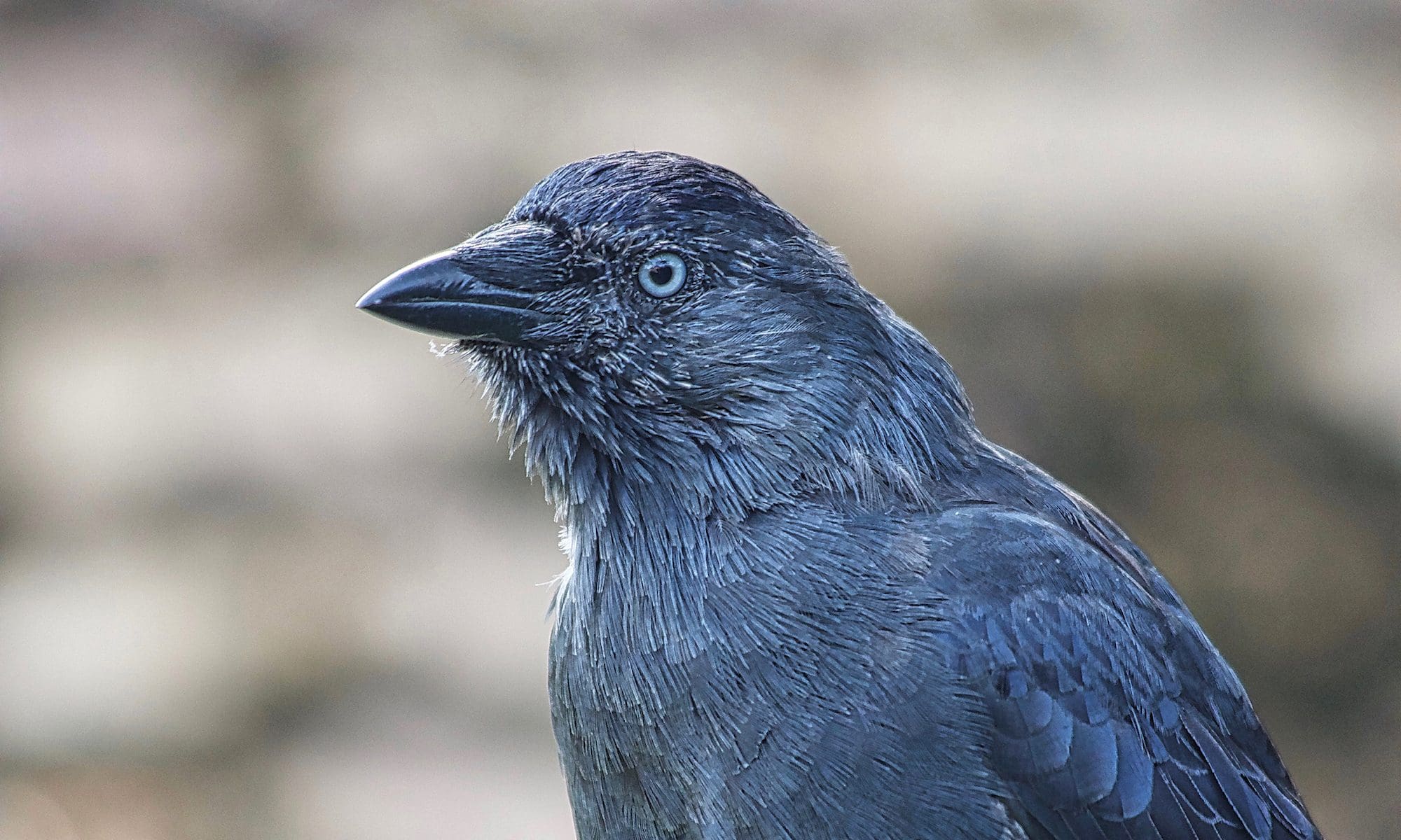
Pododermatitis, commonly known as “bumblefoot”, has become a frequently seen disease in companion and aviary birds. Pododermatitis is a general term for any inflammatory or degenerative condition of the avian foot. Pododermatitis may occur in any avian species, but is particularly problematic in permanently and temporarily captive birds, such as birds of prey, chickens, turkeys, ducks, geese, swans, waders, seabirds as well as canaries, finches, budgerigars and cockatiels.

The source of infection resulting in a pododermatitis is often a focal injury, such as a skin puncture of the undersurface of the foot through a talon, thorn or another foreign object. The second most common entry point resulting into an infection are pressure sores on the bottom of the foot. The root causes in almost all cases are the conditions animals are being kept in captivity, such as inappropriate size of cages or aviaries, inappropriately sized or textured perches or surfaces, poor nutrition and subsequent deficiencies resulting for example into obesity, unsanitary living conditions, previous mechanical or thermal injuries, inherited or acquired deformities including overgrown toenails as well as general lack of exercise and stimulation including the inability or the lack of opportunity to fly.
All these factors are contributing to a varying degree to the development of damage or focal injury causing lesions such as cracks or pressure sores to develop on the plantar surface of the phalanges or on the tarsometatarsus. The typical symptoms range from early signs, such as subtle shiny and red areas, over swollen red sores, ulcers, cuts and abrasions, to dark necrotic circular scabs. The affected bird may suffer subsequently of progressive lameness and swollen joints resulting into a reluctance to bear weight, walk, stand and grasp. If one foot is affected, a vicious cycle sets in and may affect very much soon the other healthy foot, mainly due to an altered weight distribution, which occurs as soon as the bird is trying to reduce the weight normally put onto the primarily affected foot.
Caring For Temporarily And Permanently Disabled Birds

Clinical Grades of Pododermatitis 1
Grade I: Desquamation (shedding of the epithelial or skin tissue) of small areas of the plantar foot surfaces is represented clinically by the appearance of small, shiny pink areas and peeling or flaking of the skin on the legs and feet. Initial lesions are recognised as hyperaemia (excessive amount of blood). Flattening of the skin of the digital and metatarsal pads is visible. These are the sites of maximum weight-bearing. Thinning of the plantar surface of the foot with some reddening.
Grade II: These lesions progress if untreated, and bacteria invade the subcutis, resulting in a scab and mild swelling. The subcutis is the deeper layer of the dermis, containing mostly fat and connective tissue. Smooth, thinly surfaced, circumscribed areas appear on the plantar metatarsal pads of one or both feet with the subcutaneous tissue almost visible through the translucent skin. No distinct ulcers are recognised. The thinning of the plantar surface of the foot has progressed to the point that subcutaneous tissue such as tendons can be seen through the skin.
Grade III: The sores progress to form a caseous (having a cheese-like texture) abscess with marked swelling and pain. Ulceration of the plantar metatarsal pads occurs, and in some birds, a peripheral callus may form. Ulcers form on the soles of feet with callouses forming around the edges of the lesions. Some pain and mild lameness are present.
Grade IV: Infection of the tendon sheaths develops. Corresponding cellulitis tracks toward the intertarsal joint and the digits along with flexor tendon rupture. There is a necrotic plug of tissue present in the ulcer. Most species with ulcers and accumulation of necrotic debris exhibit pain and mild lameness. Necrosis refers to cell death; the tissue turns black.
Grade V: Swelling and oedema (cellulitis) of the tissues surround the necrotic debris. The digits of the foot may also be oedematous (holding fluid). Necrotic debris starts to accumulate in the metatarsal area, indicating infection of the tendon sheaths. Severe lameness is common, and the entire metatarsal pad may be affected. This is generally a chronic lesion leading to osteoarthritis and septic arthritis of the tarsometatarsal-phalangeal joints. Cellulitis surrounds the area of necrosis, and the foot can be swollen with fluid. Tendon and metatarsal pads become infected; pain and severe lameness are present.
Grade VI: Necrotic tendons are recognised clinically as the digits swell and the flexor tendons rupture. Ankylosis and nonfunctioning digits usually present in recovery. Ankylosis refers to the stiffness of a joint due to abnormal adhesion and rigidity of the bones of the joint, which is usually the result of injury or disease. The digits are swollen and the necrotic flexor tendons on the plantar surface of the foot rupture. Even with treatment, non-functioning digits and joint fusion will be present.
Grade VII: Osteomyelitis develops. This is a bacterial bone infection leading to the destruction of the bone itself. Bone infection can progress to systemic infection and death.
Please note that all information provided in this blog is for informational purposes only and is not intended as a substitute for advice from your veterinary surgeon, physician, herbologist or other health care professional. You should not use the information on this web site for diagnosis or treatment of any health problem. Please always consult with a veterinary surgeon or healthcare professional before starting any new supplements or diet, before taking or applying any new medication, or if you suspect that your animal patient or you might have a health problem.
Even the smallest deficits in animal husbandry conditions need to be identified and rectified as soon as possible, as they may slow down or even prevent healing of injuries, or lead to recurrence after treatment. Treatment options and considerations depend on the severity of the condition. If possible, at least in early stage cases, it is suggested, if possible at all, to allow the bird to fly, as it is generally beneficial and increases blood circulation in the feet and will therefore aid healing. Sea and water birds may also benefit from being able to get back into the water, which will reduce the strain onto both feet. However, appropriate precautions need to be taken to avoid further wound infections by applying barrier ointments, waterproof bandages and by maintaining strict water hygiene measures.

For early grade lesions, improving husbandry conditions may be sufficient to prevent further deterioration and allow healing, including the thorough cleaning of perches to reduce the risk of infection. Regular prophylactic treatment of both feet with a barrier ointment such as F10 (© F10 Products Limited) should be considered. Massaging the feet whilst applying the ointment will also improve blood circulation and keep the skin in good condition. Wearing gloves should be considered to reduce the risk of transferring skin commensal bacteria to potential lesions.
If an infection is already present, topical and systemic antibiotics should be administered. Ideally, these should be based on culture and sensitivity results. Suggested options include amoxicillin / clavulanic acid or marbofloxacin. Systemic antibiotics should be continued for one week as the bare minimum, but are most likely required for longer. The wound needs to be thoroughly cleaned prior to the application of topical antibiotics. Soaking the affected foot in a dish filled with body warm water (37°C) with Epsom Salt is a commonly recommended option. Subsequent flushing of wounds with body warm hydrogen peroxid or sterile normal saline (0.9%) solution is also recommended, in particular for up to 48 hour post surgical intervention. Wounds with excessive exudate, necrotic tissue or debris may require antimicrobial cleansing solutions, which have to be non-toxic to avoid damaging healthy cells whilst encouraging wound healing.

The production of excudate (inflammatory phase of wound healing) is the body’s way of cleansing the wound and flushing all debris and bacteria inside it. At that point the wound looks pinkish or reddish, which is a sign that blood vessels are carrying oxygen-rich blood cells to nourish the wound site. This phase commonly lasts up to a week, and during this time, white blood cells are combating all bacteria to prevent an infection. Wound cleansing at this stage is important, but can also interrupt the healing process by damaging new tissue formation or reducing the temperature of the wound bed, and should therefore be done with appropriate consideration and stopped as soon as exudate production has decreased or ceased, and signs of wound healing are detected. The cleaned and dried wound area should then be treated with a topical ointment and subsequently covered, padded and dressed.
After the inflammation phase ends, the body begins to form new tissues. This phase can take a long time and usually lasts for about three weeks for minor wounds, but will be significantly longer for deeper wounds. During this phase, the body repairs damaged skin cells and blood vessels. All damaged cells are replaced with new healthy ones. These inner tissues will be covered by thin layer of skin, and the skin will pull inward causing the wound surface area to become smaller. At this point the main focus of wound care is to keep the healing wound clean and protected.

When the growth of tissues and new skin layers is complete, the next wound healing stage begins. This is the longest period and may last for months or even years. The wound site, which has healed, turns into a light red coloured wound and stretches out. The peripheral wound aspects may also start to dry out. Normal skin care and protection is recommended at this stage.
As the contralateral foot often becomes affected due to increased weight bearing, it should therefore be monitored very closely. One should also consider the use of protective bandages including barrier ointment massages on this foot to reduce the risk of pododermatitis lesions developing. Affected birds should be kept at all times on a very soft substrate such as softly padded block perches or thick towels, which need to be kept spotlessly clean. Birds, who do not move a lot around, might appreciate a soft washable u-shaped travel neck cushion, where the bird can rest on with their body and wings whilst taking the pressure off the lower extremities. A thick towel rolled into a roll and shaped accordingly can achieve a similar effect, and might actually be a bit more flexible in terms of the size required.

In more severe lesions with deep infection and granulomatous material, surgical debridement might become necessary. For these cases, protective dressings after surgery should be used to protect the wounds during healing, which includes a regular change of these bandages. One should be careful not to apply dressings too tightly, as this can potentially further compromise the blood supply to the foot. In severe cases, a hydroactive dressing might be beneficial, such as a hydrocolloid dressing, which should be applied topically to enhance wound healing. Alternatively, aloe vera gel can be used, as it helps cellular regeneration and causes the body to produce collagen. Aloe vera can help retain skin moisture and integrity while easing inflammation and preventing ulcers.
Bandaging the feet with sufficient padding to reduce and evenly distribute pressure on the plantar surfaces is required in most cases, by using for example an interdigital bandage, ball bandage or custom-fitted silicone or gel shoe, which ideally contains a central hole located precisely over the pressure sore to make sure that this area is not weight bearing at all. These dressings may require daily renewal to begin with, but will need to be changed less frequently with progressive wound healing.

Pain relief is generally recommended, and therefore analgesics such as Metacam (© Boehringer Ingelheim), which is a non-steroidal anti-inflammatory drug (NSAID), should be considered. If there is radiographic evidence of bone involvement such as osteomyelitis or bone necrosis, then the prognosis is very poor and euthanasia might be the only option left.
For more detailed information about pododermatitis and its treatment options, please follow the link below to download this excellent and comprehensive article. 2
Pododermatitis (Bumblefoot): Diagnosis, Treatment, and Resolution by J. Miesle